
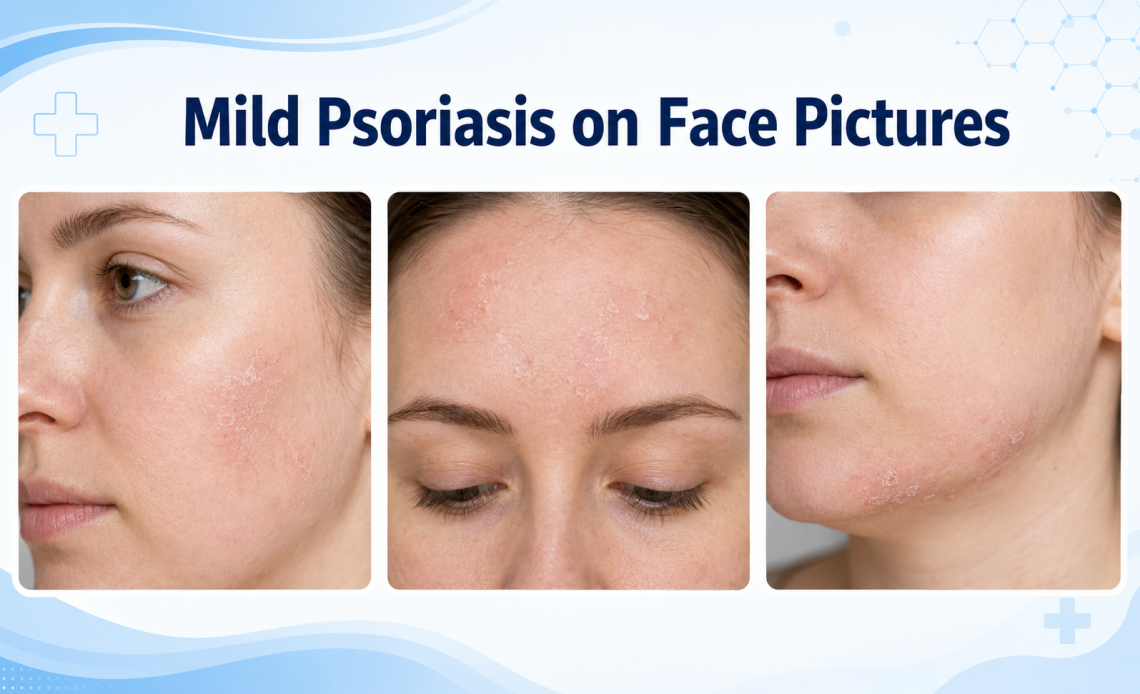
What mild facial psoriasis can look like, where it appears, how it differs from eczema and seborrheic dermatitis, and why facial treatment needs extra care
| Visual clue | Mild facial psoriasis | Common look-alike |
| Borders | Clearly defined pink, red, purple, or brown patches | Eczema often blends gradually into nearby skin |
| Scale | Fine white, gray, or silvery flaking | Seborrheic dermatitis can look greasy or yellow |
| Common sites | Hairline, eyebrows, upper forehead, around ears, and beside the nose | Contact dermatitis follows exposure to a product |
| Sensation | Itching, burning, tightness, or mild soreness | Rosacea often causes flushing and visible vessels |
| Other clues | Scalp plaques, elbow or knee patches, or nail pitting | A fungal rash may have an advancing outer edge |
Key Takeaways
- Mild facial psoriasis often appears as small, sharply defined patches with fine scale near the hairline, eyebrows, ears, or sides of the nose.
- On Black and brown skin, active patches may look purple, dark brown, gray, or ashen rather than bright red.
- Photos can help you recognize a pattern, but eczema, seborrheic dermatitis, contact dermatitis, and fungal infections can look similar.
- Facial skin is thin, so strong body-strength steroid creams should not be applied without medical guidance.
- Eye-area symptoms, rapidly spreading redness, pus, fever, or severe pain need prompt evaluation.
People searching for mild psoriasis on face pictures are usually trying to decide whether a small flaky patch is psoriasis or another common facial rash. Mild psoriasis often looks like a clearly bordered area of pink, red, purple, or brown inflammation with fine white or gray scale. It commonly appears at the hairline, between or above the eyebrows, around the ears, or in the creases beside the nose.
Pictures are useful for learning typical patterns, but they cannot confirm a diagnosis. Lighting, makeup, scratching, skin tone, previous treatment, and the location of the patch can change its appearance. A dermatologist may need to examine the scalp, nails, elbows, knees, and other body areas before deciding that the facial rash is psoriasis.
What Does Mild Facial Psoriasis Look Like?
Mild disease may involve only one or two small areas. The surface can be dry and flaky without the thick plaques usually shown in photographs of elbow or knee psoriasis. Scale is often thinner because facial skin is delicate and because moisturizers, washing, and natural skin oils reduce buildup.
A patch may itch, sting, burn, or feel tight. The border is often easier to trace than the edge of eczema. Some people also have scalp psoriasis that extends just beyond the hairline, producing a narrow band of scale on the forehead or behind the ears.
| Location | Typical appearance | Common confusion | Extra clue |
| Hairline and forehead | Fine scale extending from scalp onto skin | Dandruff or seborrheic dermatitis | Thicker scalp plaques or scale behind ears |
| Eyebrows | Small flaky, sharply bordered patches | Seborrheic dermatitis | Psoriasis elsewhere or nail pitting |
| Sides of nose | Thin red, purple, or brown patches | Rosacea or seborrheic dermatitis | Sharper borders and recurring scale |
| Eyelids | Fine scale, redness, or irritation | Eczema or allergy | Needs clinician-guided treatment |
| Around ears | Cracking or scale in front of or behind ear | Contact dermatitis | Scalp involvement is common |
| Facial psoriasis can be mild in size but high in impact
Even a small patch can affect confidence, makeup use, shaving, sleep, and social comfort. Dermatologists consider the location and effect on daily life, not only the percentage of skin involved. |
How It May Look on Different Skin Tones
On lighter skin, inflammation may appear pink or red with white or silvery scale. On deeper skin tones, the active area may look violet, deep brown, gray, or darker than the surrounding face. Scale may appear gray or ashen rather than bright white.
After inflammation improves, lighter or darker marks can remain for weeks or months. These post-inflammatory color changes do not necessarily mean the psoriasis is still active. New swelling, warmth, pus, or increasing pain is more concerning for infection.
Psoriasis Versus Eczema and Other Look-Alikes
Facial eczema is more common than facial psoriasis and often causes intense itching with less sharply defined borders. The skin may crack, ooze, or crust during a stronger flare. Psoriasis is generally more likely to form a distinct plaque and may burn or sting as much as it itches.
Capital Health Hopewell’s picture guide to eczema and psoriasis compares facial, scalp, hand, leg, and skin-fold patterns. It notes that facial psoriasis commonly affects the eyebrows, forehead, and hairline, while facial eczema often involves the eyelids, cheeks, lips, or skin around the mouth.
| Condition | Border and scale | Typical sensation | Common location |
| Psoriasis | Sharp border with fine or silvery scale | Itch, burn, or sting | Hairline, eyebrows, ears |
| Eczema | Less-defined dry patch; may ooze or crust | Often intensely itchy | Eyelids, cheeks, mouth area |
| Seborrheic dermatitis | Greasy or yellowish flakes | Itch and irritation | Scalp, eyebrows, sides of nose |
| Contact dermatitis | Matches product or exposure area | Itching or burning | Cosmetic, mask, fragrance, or hair-product contact |
| Ringworm | Expanding scaly edge, sometimes central clearing | Itchy | Any exposed facial area |
A second Capital Health Hopewell psoriasis and eczema comparison by appearance and location explains why a clinician should evaluate rashes on the face or eyelids, where thin skin changes which medications are appropriate and where fungal, allergic, and inflammatory rashes can overlap.
What Causes Facial Psoriasis Flares?
Psoriasis is an immune-mediated disease that speeds skin-cell production. A flare may follow stress, an infection, cold or dry weather, smoking, alcohol use, a skin injury, irritating products, or changes in certain medicines. Not everyone has the same triggers.
The National Institute of Arthritis and Musculoskeletal and Skin Diseases explains that psoriasis is chronic, can cycle between flares and quieter periods, and may be triggered or worsened by infections, some medicines, smoking, and other environmental or health factors. Read the NIAMS psoriasis overview for information about symptoms, causes, associated conditions, and treatment categories.
How Mild Facial Psoriasis Is Treated
Treatment aims to reduce inflammation without damaging thin facial skin. A clinician may recommend a low-potency topical corticosteroid for a short period, a steroid-sparing calcineurin inhibitor such as tacrolimus or pimecrolimus, a vitamin D-related medicine, or another prescription topical selected for the exact location.
The American Academy of Dermatology and National Psoriasis Foundation guidelines support topical corticosteroids for plaque psoriasis and identify calcineurin inhibitors and other steroid-sparing agents as useful options, particularly where steroid side effects are a concern. Review the AAD psoriasis treatment guideline Facial and eyelid treatment should be guided by a clinician because potency, duration, and product choice matter.
| Treatment type | Possible role | Important caution |
| Fragrance-free moisturizer | Reduces dryness, tightness, and visible flaking | Choose a simple nonirritating formula |
| Low-potency steroid | Short-term control of inflammation | Overuse may thin skin or affect the eye area |
| Calcineurin inhibitor | Steroid-sparing option for face or folds | May sting initially and requires a prescription |
| Other prescription topical | May control chronic facial plaques | Use only on areas directed by the prescriber |
| Systemic treatment | For psoriasis that is widespread or high impact | Requires medical screening and follow-up |
What You Can Safely Do at Home
- Wash with lukewarm water and a gentle, fragrance-free cleanser.
- Apply a bland moisturizer while the skin is slightly damp.
- Avoid scrubs, exfoliating acids, fragranced oils, and harsh acne treatments on the patch.
- Use broad-spectrum sunscreen that does not sting or trigger irritation.
- Track new cosmetics, hair products, masks, shaving products, stress, illness, and weather changes.
- Do not use a strong body steroid, coal tar, or concentrated salicylic acid on the face without medical guidance.
When to See a Dermatologist
- The rash is on the eyelids or close to the eyes.
- It has not improved after two to four weeks of gentle care.
- The diagnosis is uncertain or treatment makes it worse.
- There is pus, fever, increasing warmth, swelling, or severe pain.
- Psoriasis is affecting sleep, confidence, work, or social life.
- You develop swollen, stiff, or painful joints, heel pain, or persistent back pain.
| Get urgent help for eye or severe skin symptoms
Seek prompt medical care for eye pain, vision changes, severe eyelid swelling, a rapidly spreading blistering rash, fever with widespread redness, or facial swelling with breathing difficulty. |
Frequently Asked Questions
Can facial psoriasis look like dry skin?
Yes. Mild plaques may have only fine flaking and slight discoloration. Recurrent sharp borders, scalp involvement, and patches elsewhere make psoriasis more likely.
Does facial psoriasis always have silver scales?
No. Scale may be fine, white, gray, or barely visible, especially on oily areas or deeper skin tones.
Can I use hydrocortisone on my face?
A clinician may recommend a low-potency steroid for a short period, but facial use should be limited and guided because prolonged use can thin the skin and cause eye-area complications.
Is facial psoriasis contagious?
No. Psoriasis is immune-mediated and cannot spread through touching, towels, makeup, or close contact.
Can makeup be used over psoriasis?
Often yes when the skin is not cracked or infected. Choose fragrance-free products, patch-test first, remove makeup gently, and stop if burning or worsening occurs.
Bottom Line
Mild psoriasis on the face often appears as small, clearly defined patches with fine scale near the hairline, eyebrows, ears, or sides of the nose. On darker skin, the patch may look purple, brown, gray, or ashen instead of red. Photos can suggest a pattern, but eczema, seborrheic dermatitis, contact allergy, rosacea, and fungal infection may look similar.
Because facial and eyelid skin is thin, treatment should be gentler than treatment used on elbows, knees, or the scalp. A dermatologist can confirm the diagnosis and choose a short-term steroid or steroid-sparing medicine that is appropriate for the exact area.
| Medical disclaimer
This article provides general education and cannot diagnose a facial rash from pictures. Seek evaluation for persistent, painful, infected, rapidly spreading, or eye-area symptoms. |