
The Short Answer
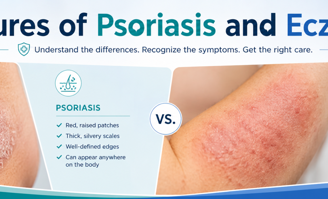
Psoriasis and eczema both cause red, inflamed, itchy skin, but they look distinctly different on close inspection. Psoriasis produces thick, well-defined plaques with a dry silvery or white scale on top, most commonly on the elbows, knees, scalp, and lower back. Eczema produces softer, less defined patches of red, weeping, or crusted skin, most commonly in the creases of the elbows and knees, on the face, and on the hands. The clearest visual distinction: if the border of the patch is sharp and the scale is thick and silvery, lean toward psoriasis. If the rash looks raw, weepy, or indistinct with intense itch, lean toward eczema. Neither can be definitively diagnosed from appearance alone. A dermatologist can confirm with a clinical exam and, when needed, a skin biopsy.
Why These Two Conditions Are So Often Confused
When people search for pictures of psoriasis and eczema, they are usually standing in front of a mirror trying to figure out which one they or their child might have. It is an understandable first step, because the two conditions can look almost identical in certain presentations, yet they are driven by different underlying mechanisms, affect different body locations, and respond to different treatments. Getting the distinction wrong means using the wrong product at best, and delaying appropriate care at worst. Psoriasis affects approximately 7.5 million adults in the United States while eczema (atopic dermatitis) affects up to 31 million Americans, making them two of the most prevalent chronic skin conditions in the country. This detailed visual and clinical guide is designed to help you identify the key differences in how each condition looks, where it appears, how it feels, and when it is time to see a specialist. For patients in the Central New Jersey area seeking a dermatology referral or skin evaluation, the clinical team at Capital Health Hopewell can connect you with appropriate specialist care.
This guide describes in precise visual terms what each condition looks like across body locations, skin tones, and severity levels, along with the key differences in sensation, triggers, and progression that help distinguish them even when appearance overlaps.
Psoriasis vs Eczema: Visual and Clinical Overview
| Feature | Psoriasis | Eczema (Atopic Dermatitis) |
| Patch appearance | Thick, raised, well-defined plaques with dry silvery or white scale | Softer, less defined rashes; may be red, weeping, crusted, or thickened |
| Border clarity | Sharp, clearly defined edges | Blurry, poorly defined edges that blend into surrounding skin |
| Scale texture | Thick, dry, flaky silvery or white scale that can be lifted or scratched off | Thin or absent scale; skin may appear raw, moist, or leathery |
| Skin color (light skin) | Red to salmon-pink base with white or silvery scale on top | Red to pink inflamed patches, sometimes with yellow crusting if infected |
| Skin color (dark skin) | Purple, gray, or ashen plaques with scale that may appear lighter or white | Darker brown, purple, or ashen patches; may be harder to detect redness |
| Common locations | Elbows, knees, scalp, lower back, nails, palms, soles | Inner elbows, back of knees, face, neck, hands, feet, wrists |
| Itch quality | Moderate itch; often described as burning or stinging more than pure itch | Intense, relentless itch, often worse at night; scratching worsens the rash |
| Skin thickness | Thick, hardened plaques that may crack and bleed | Skin may be thin and fragile or thickened and leathery from chronic scratching |
| Weeping or oozing | Rare; dry condition | Common during flares; may ooze clear or yellowish fluid |
| Nail involvement | Common: pitting, ridging, yellow-brown discoloration, nail lifting | Occasional: ridging, thickening from scratching |
| Joint symptoms | Psoriatic arthritis in up to 30 percent of patients | Not directly associated with joint disease |
What Psoriasis Looks Like: A Detailed Visual Description
Plaque Psoriasis: The Most Common Type
Plaque psoriasis accounts for approximately 80 to 90 percent of all psoriasis cases. The defining visual feature is a raised, thickened patch of skin called a plaque, with a dry, adherent scale on top. On lighter skin tones, the base of the plaque is typically a deep red or salmon-pink color. The scale overlying it is white to silvery in color and has a distinctive dry, almost waxy texture. When the scale is gently removed, it often reveals fine bleeding points underneath, a clinical sign called the Auspitz sign, which is specific enough to psoriasis that dermatologists use it as a diagnostic clue.
The plaques have sharply defined, clearly visible edges where the affected skin meets normal skin. This clarity of border is one of the most reliable visual distinctions from eczema. Plaques range from the size of a small coin to large confluent areas covering the elbows, knees, or scalp entirely. The skin within the plaque is dry, never weeping, and may develop painful cracks (fissures) particularly on the palms, soles, and over knuckles.
Psoriasis on the Scalp
Scalp psoriasis is one of the most common and most mistaken presentations of the condition. It produces thick, silvery-white scale on the scalp that can extend just beyond the hairline onto the forehead, behind the ears, and on the back of the neck. This scale is denser and more adherent than the flaky dandruff of seborrheic dermatitis, and the underlying scalp is red and inflamed. Hair loss is not a direct result of psoriasis but can occur from repeated trauma from scratching.
Psoriasis on the Nails
Nail psoriasis is present in approximately 50 percent of people with psoriasis and in almost all people who have psoriatic arthritis. The most characteristic finding is pitting, small depressions on the nail surface that look as if a pin has been pressed into the nail repeatedly. Other nail changes include a brownish-yellow discoloration under the nail called oil drop sign, onycholysis (separation of the nail from the nail bed), and subungual hyperkeratosis, a chalky white buildup under the nail plate. Nail changes in psoriasis are often misidentified as fungal nail infection.
Psoriasis on Dark Skin Tones
Psoriasis presents differently on darker skin tones and is frequently underdiagnosed or misidentified as a result. Rather than the vivid red plaques seen on lighter skin, psoriasis on brown or Black skin tends to appear as purple, violet, gray, or ashen plaques. The scale may appear lighter or white against a darker skin background. The plaques are still raised and well-defined, and the Auspitz sign (pinpoint bleeding when scale is removed) remains a useful clinical marker regardless of skin tone. Post-inflammatory hyperpigmentation, where dark marks persist in areas of healed psoriasis, is more prominent and longer-lasting on darker skin.
Types of Psoriasis and How They Look
| Type | Appearance | Common Locations |
| Plaque psoriasis | Raised, thick, silvery-scaled plaques with sharp borders | Elbows, knees, scalp, lower back |
| Guttate psoriasis | Small (0.5 to 1.5 cm), drop-shaped red spots, often following strep infection | Trunk, arms, legs; often sudden onset in children and young adults |
| Inverse psoriasis | Smooth, shiny, bright red patches without scale; skin-on-skin friction aggravates | Skin folds: armpits, groin, under breasts, around genitals |
| Pustular psoriasis | White or yellow pus-filled blisters surrounded by red, inflamed skin | Palms and soles (palmoplantar), or widespread across the body |
| Erythrodermic psoriasis | Widespread fiery redness covering most of the body; skin peels in sheets | Entire body; rare, severe, medical emergency |
| Nail psoriasis | Pitting, oil-drop discoloration, onycholysis, subungual hyperkeratosis | Fingernails and toenails |
What Eczema Looks Like: A Detailed Visual Description
Acute Eczema Flare
During an active flare, eczema produces intensely red, swollen skin that may weep clear or slightly yellowish fluid. The skin surface looks raw and wet, sometimes forming small fluid-filled blisters called vesicles that break and crust over. This weeping, crusted appearance is highly characteristic of eczema and is virtually never seen in plaque psoriasis. The borders of the rash are indistinct, fading gradually into the surrounding normal skin rather than ending with the sharp edge seen in psoriasis.
Chronic Eczema
With repeated scratching over months and years, eczema-affected skin undergoes a process called lichenification, where the skin becomes thick, leathery, and deeply grooved from the chronic mechanical trauma. Lichenified eczema can look superficially similar to a psoriatic plaque because both are thickened and raised, but the texture is different: lichenified eczema has an accentuated skin line pattern and is often darker than surrounding skin, while psoriatic plaques have the characteristic silvery scale. Lichenified eczema patches also tend to be in the flexural creases (inner elbows, back of knees) rather than the extensor surfaces where psoriasis predominates.
Eczema by Body Location
| Location | How Eczema Appears There | Distinguishing Feature |
| Inner elbows and back of knees | Red, scaly, sometimes weeping patches in the crease; may be lichenified in chronic cases | Flexural location is classic for eczema; psoriasis typically appears on outer surfaces |
| Face (especially cheeks in children) | Red, dry, rough patches on cheeks and around the mouth in infants and toddlers | Facial eczema is common in childhood; facial psoriasis more common in adults |
| Eyelids | Swollen, red, scaly thin skin; eyelid skin is very thin and reacts intensely | Eyelid involvement more common in eczema than psoriasis |
| Hands and wrists | Red, dry, cracked skin on backs of hands; vesicles on palms in dyshidrotic eczema | Dyshidrotic eczema (tiny blisters on palms and soles) is unique to eczema |
| Neck | Red, irritated skin that may be lichenified; often worse with sweat and friction | Neck involvement very common in eczema from contact with fabrics and hair products |
| Nipples | Dry, scaly, irritated skin around the nipple area | Nipple eczema is common; nipple psoriasis less so but exists |
Eczema on Dark Skin Tones
Eczema is more prevalent in people with darker skin tones, and it presents differently. Rather than the vivid redness of eczema on lighter skin, eczema on brown or Black skin may appear as darker brown, purple, or grayish patches. The skin may also show follicular accentuation, where the hair follicles become prominent, giving the skin a rough, goosebump-like texture that is less common in lighter skin presentations. Post-inflammatory hyperpigmentation leaves dark marks at previous eczema sites that can persist for months after the active rash resolves.
The single most reliable visual rule for telling them apart at a glance: psoriasis appears on the outside of joints (elbows, knees) with a sharp silvery scale. Eczema appears inside the joints (inner elbow crease, back of knee) with a blurry, raw, or weeping appearance. Location plus scale type gets you most of the way to the right diagnosis before a doctor even examines the skin.
Different Causes, Different Triggers
| Factor | Psoriasis | Eczema |
| Root cause | Autoimmune: immune system attacks skin cells, causing them to multiply 10x faster than normal | Skin barrier dysfunction plus immune overreaction to environmental triggers |
| Genetic component | Strong: 10 percent risk if one parent affected; 50 percent if both parents affected | Strong: 80 percent of people with eczema have a family history of eczema, asthma, or hay fever |
| Common triggers | Stress, strep infection, skin injury (Koebner phenomenon), certain medications (lithium, beta-blockers), alcohol | Soaps, detergents, wool, pet dander, dust mites, sweat, dry air, food allergens (in children), stress |
| Infectious trigger | Guttate psoriasis triggered by streptococcal infection | Eczema flares triggered or complicated by Staphylococcus aureus skin colonization |
| Age of typical onset | Peaks at ages 15 to 35 and 50 to 60 | Usually begins in childhood; 60 percent of cases before age 1 |
How Treatments Differ Between the Two Conditions
Because psoriasis and eczema have different underlying mechanisms, their treatments overlap in some areas but differ importantly in others. Using the wrong treatment can be ineffective or even counterproductive.
- Topical corticosteroids are used for both conditions and are often the first-line treatment for mild to moderate cases of each. However, the strength of steroid required and the body location where it is safe to use differ between conditions.
- Moisturizers are central to eczema management and less critical for psoriasis. Eczema involves skin barrier dysfunction that is directly improved by consistent, heavy emollient application. Psoriasis plaques benefit from moisturizers to reduce scale and discomfort, but moisturizer alone does not address the underlying immune process.
- Biologic medications target different immune pathways for each condition. For psoriasis, biologics targeting IL-17, IL-23, and TNF-alpha are highly effective. For eczema, dupilumab (Dupixent), which targets IL-4 and IL-13, is the most widely used biologic and was specifically developed for atopic dermatitis.
- Coal tar preparations are used for psoriasis but are generally not used for eczema. Salicylic acid in shampoos and topicals is used to reduce psoriasis scale but is not indicated for eczema.
- Phototherapy (UV light treatment) is effective for both conditions but is more commonly used as a standard treatment escalation in psoriasis.
When to See a Dermatologist
Many people with mild psoriasis or eczema manage their condition successfully with over-the-counter products and trigger avoidance for months or years before seeking specialist evaluation. According to Medical News Today, a dermatologist should be consulted when the condition is unresponsive to over-the-counter treatments, covers a significant body surface area, affects the face, hands, or genitals, causes sleep disruption, or has an uncertain diagnosis. The following specific situations warrant prompt medical evaluation:
- Any skin rash that has not improved after two to four weeks of consistent over-the-counter treatment
- A rash covering more than 10 percent of the body surface area
- Signs of skin infection at a rash site: increased warmth, swelling, pus, or fever
- Joint pain or swelling in someone with a known or suspected psoriasis diagnosis, as this may indicate psoriatic arthritis requiring separate treatment
- A new rash in an infant or young child, to confirm the diagnosis and establish a safe treatment plan
- Any rash on the eyelids, face, or genitals, where skin is thin and requires specialist guidance on treatment selection
- Rash that appears suddenly across a large area of the body, particularly following an illness or new medication
The Visual Detail That Confuses Even Experienced Observers
The overlap zone where psoriasis and eczema are genuinely difficult to distinguish visually is the hand. Both conditions produce dry, cracked, inflamed skin on the palms and fingers. The key differentiator that dermatologists use is the pattern of fissuring and the presence or absence of scale type. Psoriasis on the hands tends to produce uniform, thickened plaques with white scale over the entire palm, while eczema of the hands often shows patchy, asymmetric involvement with intense itch and possible vesicles (tiny blisters) between the fingers or on the lateral palm, a subtype called dyshidrotic eczema that has no equivalent presentation in psoriasis.
A second visual detail worth knowing: inverse psoriasis, which affects skin folds like the armpit, groin, and under the breasts, looks nothing like classic plaque psoriasis. It produces smooth, shiny, bright red patches without any of the characteristic scale, because moisture in the folds prevents scale formation. This presentation is commonly mistaken for fungal infection (intertrigo) or eczema. The key clue is that inverse psoriasis tends to have sharper, cleaner borders than either fungal infection or eczema, and the patient typically has or has had psoriasis elsewhere on the body.
The Bottom Line
Psoriasis and eczema share enough surface-level similarities to cause genuine confusion, but their most important visual characteristics are reliably different. Psoriasis produces thick, well-defined, silvery-scaled plaques on extensor surfaces and often involves the scalp and nails. Eczema produces poorly defined, weeping or dry patches in skin creases and flexural areas, with intense itch as its dominant symptom. Location, border clarity, scale type, and itch quality are the four features that most reliably distinguish the two conditions before a diagnosis is confirmed.
A dermatological diagnosis confirmed by a trained clinician is the only definitive way to tell the two apart, particularly in atypical presentations, on darker skin tones, or when the rash does not respond to initial treatment as expected. If you are in the Mercer County or Central New Jersey area and need a clinical evaluation for a skin condition, Capital Health Hopewell can connect you with dermatology and specialist services for a confirmed diagnosis and personalized treatment plan.